Table of Contents
- Resting Phase: The Sleeping Guardians
- Injury Signals and Activation
- Proliferation: The Building Phase
- Differentiation and Fusion: Rebuilding the Muscle
- Self-Renewal: Saving a Backup
- Influencing Factors That Shape Recovery
- How does this compare to Past Understandings?
- Why It Matters?
- Future Directions in Research
- Conclusion
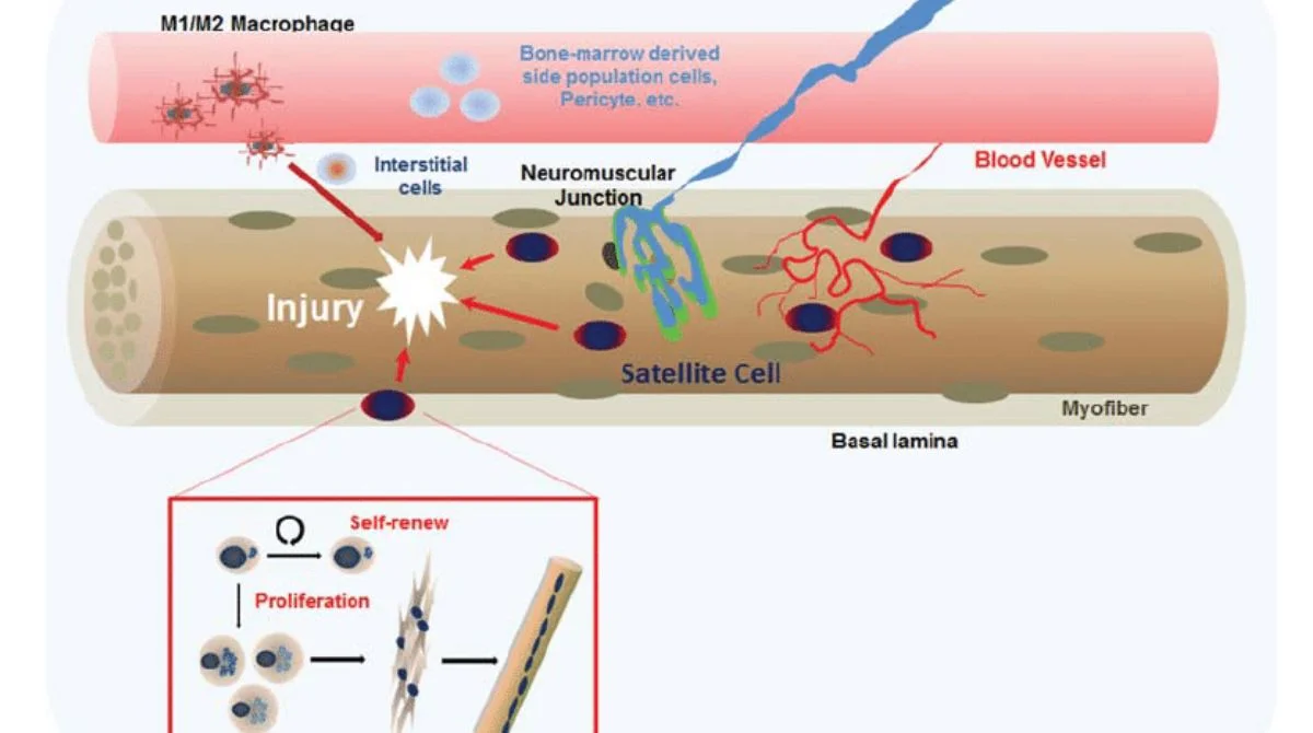
When muscles get injured through heavy lifting, a fall, or surgery, your body calls in its special repair team: myosatellite cells (also known as satellite cells or MuSCs). These tiny, mostly inactive cells lie quietly between the muscle fiber cover (sarcolemma) and the surrounding layer (basal lamina), waiting for their moment to act. In this article, we’ll walk through how these cells wake up, multiply, and rebuild muscle and why that matters for healing, fitness, and aging.
Resting Phase: The Sleeping Guardians
Under normal conditions, most myosatellite cells are asleep in a phase called G₀ quiescence. They show few cell parts, making them easy to overlook. But they’re vital, poised, and ready to respond as soon as they’re needed.
Injury Signals and Activation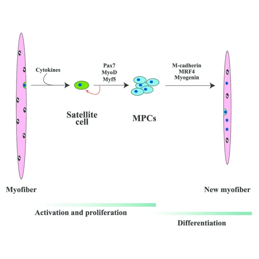
When muscle fibers are damaged, they leak proteins and change signals around them. Immune cells like neutrophils and macrophages rush in to clean up, releasing cytokines and growth factors such as HGF, IGF-1, and interleukins. These signals jolt satellite cells out of their slumber.
Two key genes, Pax7 and Pax3, help mark and turn the cells on. Pax7 is always on in active satellite cells, while Pax3 is more limited.
Proliferation: The Building Phase
Once awake, satellite cells re-enter the cell cycle and rapidly multiply, becoming a “transit-amplifying pool” of myoblasts. They aim to bring enough cells to the injury site.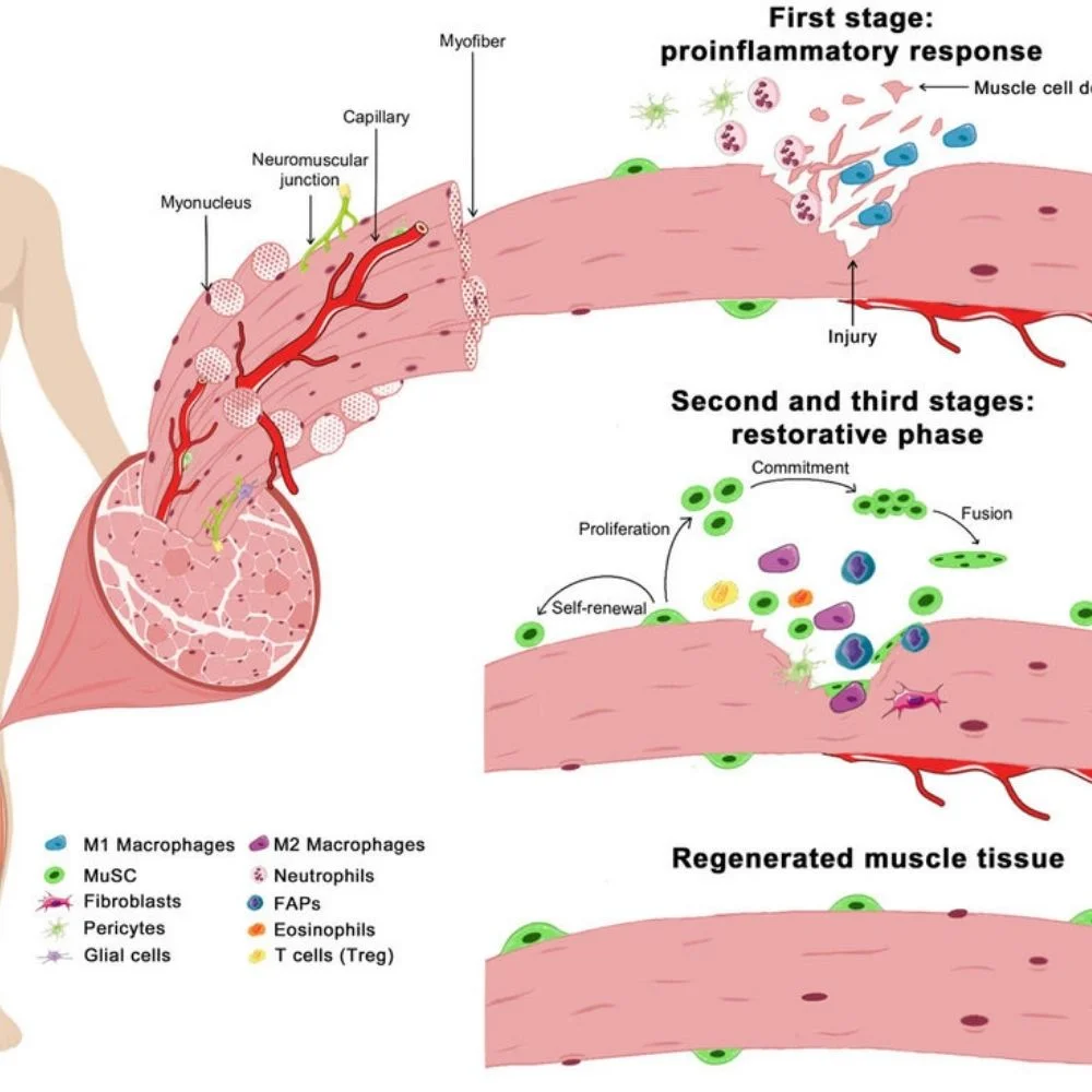 They increase the expression of muscle-related genes like MyoD, Mrf4, and myogenin, which help control which way the cells move from dividing to fusing into fibers.
Differentiation and Fusion: Rebuilding the Muscle
The myoblasts then begin to differentiate and fuse with each other or with damaged fibers to rebuild muscle structure. This process involves rebuilding the extracellular matrix (ECM), replacing scar tissue, and returning structure and strength.
A clean balance between two macrophage types, pro-inflammatory M1 (cleanup) and anti-inflammatory M2 (rebuilding), helps the process run smoothly.
Self-Renewal: Saving a Backup
Not all satellite cells get used up. Some self-renew via asymmetric division, keeping one copy to stay as a reserve and pushing another to join the repair work. This keeps the muscle’s regenerative potential alive for the future.
Influencing Factors That Shape Recovery
Several factors impact how well recovery goes:
- Growth factors: HGF triggers activation; IGF-1 supports both multiplication and fusion of cells.
- Signaling pathways: Levels of Notch, Wnt, and PI3K/Akt/mTOR guide satellite cells between multiplying, muscle commitment, or self-renewal.
- Niche cues: ECM components (like fibronectin, laminin) and nearby support cells influence decisions inside satellite cells.
- Inflammation: The right immune reaction is vital; too much or too little can confuse satellite cells.
- Age and load: With age or long rest, satellite cell numbers and performance drop. Resistance training and moderate exercise can boost their counts.
How does this compare to Past Understandings?
Earlier views treated satellite cells like simple repair workers, but new research shows how complex and regulated they are:
- We now know more about niche influence, epigenetics, and aging effects.
- We also understand how exercise alone, without actual injury, can activate satellite cells and grow muscle.
- Research highlights the need for a precise switch in signals to keep regeneration effective and scar-free.
Why It Matters?
Knowing how myosatellite cells work is key for:
- Better healing: Targeting pathways like IGF1 or NFκB to speed up recovery.
- Anti-ageing: Encouraging satellite cell reserves to slow muscle loss.
- Regenerative medicine: Building bioengineered muscles or stem cell therapies.
- Athletic recovery: Fine-tuning training and recovery strategies for faster repair.
Future Directions in Research
Next big steps in the field include:
- Mapping satellite cell diversity using single-cell studies.
- Finding ways to tweak their niche for better outcomes.
- Developing better lab models for drug testing and tissue engineering.
- Studying how space travel or zero-gravity affects them.
Conclusion
Myosatellite cells are the secret stars behind muscle repair. From their quiet beginnings, they rise to multiply, rebuild muscle fibers, and replenish themselves for future needs. With a coordinated dance of signals from immune cleanup crews, growth hormones, nearby cells, and structural scaffolding, they bring muscles back to health. Research is now untangling the deep complexity of their actions and finding ways to enhance healing, reduce scarring, and boost muscle resilience. Myosatellite cells are tucked-away muscle stem cells; when injury strikes, they wake, copy themselves, rebuild damaged fibers, and save a backup for later with signals and support that modern science is learning to optimize for faster, cleaner muscle recovery.